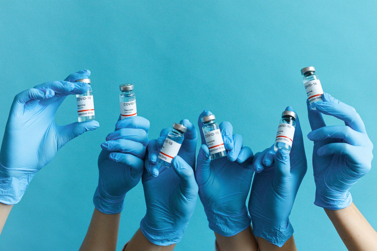
Featured coverage image. Source: Editorial desk.
Johns Hopkins researchers have uncovered that Vitamin A plays a critical role in the fetal development of sharp central vision, challenging long-held theories. This discovery offers new hope for treating degenerative eye conditions through advanced regenerative medicine.
Johns Hopkins researchers have uncovered that Vitamin A plays a critical role in the fetal development of sharp central vision, challenging long-held theories. This discovery offers new hope for treating degenerative eye conditions through advanced regenerative medicine.
Humans possess an extraordinary capacity for high-acuity, full-color central vision—a trait shared by very few mammals. Early in 2026, researchers at Johns Hopkins University shattered a decades-old scientific consensus regarding how this sharp vision develops. Published in the Proceedings of the National Academy of Sciences (PNAS), their study reveals that Vitamin A does not merely support our eyes; it actively choreographs the cellular transformation required to build the foveola, the tiny hub of high-resolution sight. This discovery challenges established textbooks and paves the way for advanced therapies targeting blindness and macular degeneration.
Historically, vision science assumed that the critical photoreceptor cells responsible for our sharpest vision migrated into place during embryonic development. The findings from the Johns Hopkins team reveal a far more dynamic, chemically driven metamorphosis. By mapping this process, scientists can now replicate human retina development with unprecedented accuracy in laboratory settings, opening new horizons for regenerative medicine.
Vision development is the precise biological sequence through which the mammalian eye, retina, and corresponding neural pathways mature to capture, process, and interpret visual stimuli. In humans, this journey involves the differentiation of specialized photoreceptors—rods for low-light navigation and cones for color and high-acuity details—culminating in the formation of the macula and foveola for sharp central focus.
The human retina is a complex, multi-layered tissue lining the back of the eye. While rods outnumber cones across the general surface of the retina, the foveola—a minuscule depression at the center of the macula—contains exclusively cone photoreceptors. These cones are further specialized to detect different wavelengths of light: red (long-wavelength), green (medium-wavelength), and blue (short-wavelength). How this tiny region becomes packed with red and green cones while excluding blue ones has remained a biological mystery until now.
The research team, led by Robert J. Johnston Jr., an associate professor of biology at Johns Hopkins University, identified a meticulously timed interaction between thyroid hormones and retinoic acid—a molecule derived from Vitamin A. This molecular crosstalk dictates the fate of photoreceptors in the developing human foveola before birth.
For decades, the leading theory posited that blue cones initially populated the center of the retina but later migrated outward to leave a pure concentration of red and green cones. The Johns Hopkins study disproved this passive migration theory. Using advanced stem cell techniques, the researchers demonstrated that blue cones do not migrate; instead, they undergo a direct cellular reprogramming, converting into red or green cones during fetal development.
This cellular conversion occurs during a narrow gestational window, specifically between weeks 10 and 14. According to Johnston, understanding this temporal window and the chemical triggers that control it is a critical step toward reconstructing the human retina. This knowledge is particularly vital for addressing macular degeneration, where the foveola is the first region to suffer irreversible damage.
The transformation of the foveola relies on a precise, two-part biochemical relay. To observe this phenomenon, the research team utilized human retinal organoids—three-dimensional tissues grown from stem cells that mimic the structure and development of the human eye in vitro.
Below is a schematic outline of this newly discovered developmental pathway:
[Weeks 10–12 of Fetal Development]
               │
               ▼
Foveola populated primarily by Blue Cone Photoreceptors
               │
               ▼
[Step 1: Retinoic Acid Degradation]
Vitamin A-derived retinoic acid levels are deliberately suppressed in the foveola,
preventing the generation of new blue cones.
               │
               ▼
[Step 2: Thyroid Hormone Escalation]
Thyroid hormones flood the foveolar tissue, triggering a genetic switch.
               │
               ▼
[Weeks 14+ of Fetal Development]
Existing blue cones reprogram and convert into Red and Green Cone Photoreceptors
This two-stage mechanism shows that retinoic acid acts as a spatial patterning agent. By degrading retinoic acid in the central retina, the body halts blue cone production in that specific zone. Subsequently, thyroid hormones act as a differentiation switch, forcing the remaining immature blue cones to adopt red or green identities.
If this process fails, blue cones remain in the foveola. Because blue light scatters more easily within the eye, the presence of blue cones in the central focal point significantly degrades visual sharpness. Thus, the local depletion of Vitamin A's derivative, followed by thyroid hormone activation, is mandatory for high-definition human vision.
By decoding the natural blueprint of the foveola, ophthalmologists and regenerative medicine researchers can improve strategies to combat retinal diseases. Conditions like Age-Related Macular Degeneration (AMD) and glaucoma cause profound visual impairment by destroying the delicate cellular architecture of the eye.
| Condition | Primary Pathology | Affected Demographics (U.S.) | Strategic Impact of the Discovery |
|---|---|---|---|
| Age-Related Macular Degeneration (AMD) | Progressive damage to the macula and foveola, destroying central vision. | Over 11 million individuals, primarily aged 60+ | Enables researchers to generate target-specific red and green cones for macular transplantation. |
| Glaucoma | Optic nerve damage often linked to intraocular pressure, leading to blindness. | Over 3 million individuals | Illuminates pathways for preserving retinal ganglion cell health and restoring neural wiring. |
The immediate beneficiary of this research is organoid technology. Previously, lab-grown retinal organoids lacked the precise arrangement of photoreceptors found in a natural human eye. By manipulating retinoic acid and thyroid hormone levels at specific development intervals, the Johns Hopkins team can now grow retinal tissues that feature a true, high-acuity foveola.
This progress shifts the medical conversation from slowing vision loss to actively restoring sight. In the future, surgeons may transplant these customized, lab-grown patches of red and green cones directly into the maculas of patients suffering from advanced AMD. Additionally, because these organoids mimic human development far more accurately than animal models, they provide an ideal testing ground for new pharmaceuticals, accelerating drug discovery while reducing reliance on animal testing.
While the 2026 Johns Hopkins study uncovers a prenatal role for Vitamin A, the nutrient's relationship with vision has been documented for thousands of years. As early as 3500 BC, ancient Egyptian physicians prescribed topically applied or consumed raw liver—now known to be an exceptionally rich source of Vitamin A—to treat night blindness.
The modern scientific understanding of Vitamin A began in 1913, when biochemist Elmer McCollum co-discovered "fat-soluble A" at the Wisconsin Agricultural Experiment Station. McCollum, who later became the first chair of the Department of Biochemistry at the Johns Hopkins School of Hygiene and Public Health, demonstrated that a lack of this nutrient caused severe ocular lesions and blindness in animal models.
Decades later, in the 1970s and 1980s, Johns Hopkins epidemiologist Alfred Sommer proved that Vitamin A supplementation could prevent xerophthalmia (dry eye disease) and reduce child mortality rates globally. Sommer’s work prompted international public health organizations to launch mass Vitamin A distribution campaigns, saving millions of children from preventable blindness.
Within the mature eye, Vitamin A is converted into retinaldehyde (retinal), which binds with proteins called opsins to form rhodopsin and iodopsin—the light-sensitive pigments inside rods and cones. When light hits these pigments, it triggers a chemical change that sends electrical signals to the brain. Without continuous dietary intake of Vitamin A, the eye cannot regenerate these visual pigments, leading to night blindness and eventual corneal degradation.
Although prenatal retinal development relies on delicate hormonal balances, maintaining adult vision requires a steady, lifelong supply of Vitamin A and other supportive micronutrients.
The discovery of how Vitamin A shapes the foveola is catalyzing further innovations across the Wilmer Eye Institute at Johns Hopkins. Researchers are combining these organoid models with CRISPR gene-editing technology to correct hereditary retinal diseases before transplantable tissues are grown.
Concurrently, scientists are tackling the challenges of optic nerve regeneration. A study published in Science Translational Medicine highlighted progress in transplanting retinal ganglion cells and encouraging them to form functional connections with the brain. By combining photoreceptor transplantation with optic nerve repair, regenerative ophthalmology is moving closer to reversing complex causes of blindness.
Vitamin A is converted by the developing fetus into retinoic acid. Scientists discovered that the degradation, or clearing out, of retinoic acid in the center of the retina during weeks 10 to 12 of pregnancy is the signal that stops the production of blue cones. This clearing step, followed by a surge in thyroid hormones, forces existing photoreceptors in the foveola to transform into red and green cones, which are required for sharp central vision.
Blue light has a shorter wavelength and scatters more easily than red or green light, which can create visual blur. If blue cones remain in the foveola—the center of our visual focus—it decreases overall visual acuity. By converting prenatal blue cones into red and green cones, the body ensures that the foveola is optimized for high-definition detail, reading, and color distinction.
Retinal organoids are three-dimensional, lab-grown mini-retinas derived from human stem cells. Because animal models like mice do not possess a foveola, researchers could not study human central vision development using traditional animal testing. Retinal organoids allowed Johns Hopkins scientists to observe and manipulate human retinal development in real-time, leading to the discovery of the retinoic acid and thyroid hormone pathway.
While adequate Vitamin A intake is necessary to maintain normal visual pigments and prevent night blindness, consuming excess Vitamin A will not alter the physical structure of your foveola or cure existing macular degeneration. Prenatal development pathways differ from adult eye maintenance. To protect your vision, aim for recommended daily allowances of Vitamin A through a balanced diet rather than high-dose supplements, which can be toxic.
Featured image by Maksim Goncharenok on Pexels
AI BlogX is committed to high editorial standards. For time-sensitive or critical topics, please verify claims against original primary sources.
Authoritative and trend-focused coverage across business, sports, entertainment, health, lifestyle, politics, science, and technology.
More Desks
© 2026 AI BlogX. All rights reserved.
Trend-focused editorial workflow

Stories are monitored from trending signals, then processed for accurate summaries, fact-checking, and desk oversight.
Editorial policy
